Fluid and electrolyte balance
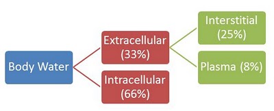
Body fluids consists of intracellular and extracellular fluid. Hydrostatic pressure and osmotic pressure is responsible for the movement of water through the various body fluid compartments. Change in the osmolality of the extracellular fluid due to ingestion of water and salt result in the movement of water between the extracellular fluid and intracellular fluid. Water will move towards the compartment with the higher osmolality to maintain equilibrium.
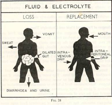
Various factors cause EC and IC volumes to change which may result in disorders, these factors include : ingestion of H20 or dehydration, IV transfusions of various solutions, loss of large amount of fluid from GIT, loss of abnormal fluids from GIT, loss of abnormal fluids though sweating or kidneys.
Sodium control mechanisms include : Glomerular Filtration Rate, Renin-Angiotensin-Aldosterone mechanism & Atrial Natriuretic Peptide.
Oedema results whenever small blood vessels increases in permeability, resulting in accumulation of extracellular fluid into nearby tissues causing tissue to swell (fig 1)

There are a number of physiological abnormalities that can result in a rise in interstitial fluid pressure and edema. Low concentration of proteins in blood plasma decreases osmotic pressure that normally acts to prevent the leakage of fluid from blood vessels into the extracellular tissue space. In the case of trauma or inflammation, there is usually an increase In the permeability of the capillary wall that results in greater fluid passage into tissue spaces. Generalized oedema include cardiac oedema, renal oedema, famine oedema, toxaemia of pregnancy. Localized oedema include Cerebral oedema, hepatic oedema, pulmonary oedema.
Hyperaemia and congestion means increased volume of blood in a particular site. Hyperaemia is an active process in which arteriolar dilation as occurs with skeletal muscle during exercise or at sites of inflammation. Tissues are red (erythema) due to engorgement with oxygenated blood.(fig 2)

Congestion is a passive process resulting from impaired outflow of blood from tissue. Tissue has an abnormal blue-red color (cyanosis) due to accumulation of deoxygenated haemoglobin. Edema is usually the result of congestion due to volumes and pressures being increased.(fig 3)

Haemorrhage Is the loss of blood due to a rupture of a vessel. (fig 4 & 5)


May range from minor injuries to life threatening emergencies. The effects of haemorrhage depends on the volume of blood lost, the rate at which its lost and the site of injury. The body responds to blood loss by redistributing the blood to vital centers.
Shock
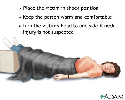
Shock can be caused by any condition that reduces blood flow, can cause damage due to a lack of oxygen to multiple organs and tissues. It is a life threatening condition if untreated due to it being a progressive disorder. “Major causes include Hypovolemic shock – internal and external bleeding, Septic shock – infections in the bloodstream, Anaphylactic shock – severe allergic reaction, Cardiogenic shock – inability of heart to pump blood effectively, Neurogenic shock – extreme emotional upset.” Treatment of shock depends on the cause.
Depending on the specific cause and type of shock, symptoms will include one or more of the following:
Anxiety or agitation, restlessness, bluish lips and fingernails, chest pain confusion, dizziness, lightheaded, pale, cool, clammy skin, low or no urine output, profuse sweating, moist skin, rapid weak pulse, shallow breathing, unconsciousness.
Thrombosis is the formation of a blood clot within an uninterrupted cardiovascular system. Thrombosis is caused due to disturbed flow of blood, hypercoagulability and injury to endothelial lining of the blood vessel. (fig 6)

Embolism is the condition when a complete or part of a blood clot detaches itself from its site and causes occlusions at another part of the body. Major cause is deep vein thrombosis. In this condition, a blood clot is formed deep inside the vessel in the thigh or lower leg.(fig 7)

References:
Rippey J.J. General Pathology 1985. Witwaterstrand University Press. JHB
Underwood J.C.E. General and Systemic Pathology, 2009, 5TH ed. Chirchill Livingstone
Fluid and electrolyte balance in the body
http://faculty.ucc.edu/biology-potter/fluid_and_electrolyte_balance_in.htm
Oedema – Family Doctor
http://www.familydoctor.co.nz/conditions.asp?A=32853&category_name=
Hyperaemia and Congestion
MedlinePlus Medical Encyclopedia
